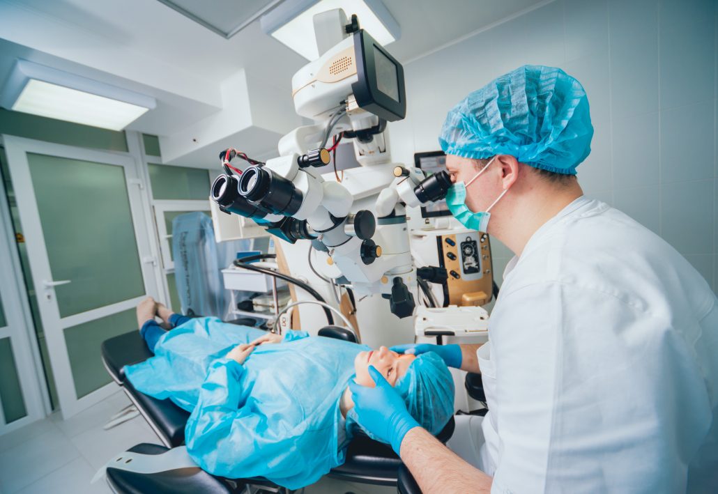
DIABETIC LASER TREATMENT
Laser photocoagulation is a proven effective treatment for preserving vision and reducing the risk of vision loss from diabetic retinopathy. In national clinical trials of laser treatment for diabetic retinopathy conducted at multiple locations, laser photocoagulation reduced the risk of severe vision loss from diabetic retinopathy to less than 2% over a five-year period.
Two laser techniques are performed today, generally using an argon laser:
- Scatter or panretinal photocoagulation generally requires 1,200-1,800 individual laser spots, usually spread over two or three sessions. In this technique the ophthalmologist avoids the macula, the central area of the retina that is responsible for our reading vision, color vision, and other tasks requiring sharp vision. Scatter laser photocoagulation is used to treat proliferative diabetic retinopathy, a major cause of severe vision loss from diabetes.
- Focal laser surgery uses fewer spots and less intense laser power to treat diabetic macular edema. Using a technique called fluorescein angiography and other examination and photographic techniques, the ophthalmologist identifies areas that are leaking fluid into the macula area. These areas are then treated directly with a laser to prevent further leakage of fluid into the macula and to allow fluid that has already leaked to be reabsorbed.

